NEOLITHIC SURGERY

3,000 year prosthetic toe
An excavation of a 6,900-year-old tomb at Butheirs-Boulancourt, about 65 kilometers south of Paris, revealed a man with an amputated forearm. To perform such an operation would require a high degree of skill and knowledge about the human body an infection. According to the French National Institute for Preventive Archaeological Research, the patient seemed to haven been anaesthetized, the conditions were aseptic, the cut was clean and the wound was treated. Scientists believe that very sharp flint tools were used to do the cutting, which included cutting through bone, and plants such as sage may have been used to clean the wounds and as an anaesthetic. See Borneo Amputation Below
There is also evidence of Neolithic amputations being performed in Germany and the Czech Republic. It had been known for some time that Stone Age men performed trephinations, cutting holes in the skull, but these are the first evidence of amputations. The elderly amputee lived during the Linearbandkeramik period, when European hunter-gatherers began settling down to agriculture stockbreeding and pottery. A schist axe, a flint pick and other iams of a young animal, thought to be a sign of high status was found in the amputee’s grave.
According to Archaeology magazine: A hole in a 5,200-year-old cow skull is evidence of Neolithic bovine brain surgery. When the cranium was originally found at Champ-Durand, France, it was thought that the hole was caused by another cow’s horns, but reanalysis confirmed that the aperture’s characteristics are more consistent with trepanation. Experts believe that perhaps the world’s first known veterinarian attempted to save the cow’s life through surgery, or that Neolithic surgeons honed their skills on domestic animals before applying them to human subjects. [Source: Archaeology magazine, July- August 2018]
RELATED ARTICLES:
HEALTH IN ANCIENT GREECE europe.factsanddetails.com ;
DISEASE IN ANCIENT GREECE factsanddetails.com ;
HEALTH CARE IN ANCIENT GREECE: TREATMENTS, HEALING TEMPLES, MIRACULOUS CURES europe.factsanddetails.com ;
DOCTORS AND HEALTH CARE PRACTITIONERS IN ANCIENT GREECE europe.factsanddetails.com ;
MEDICINES IN ANCIENT GREECE: CURES, HERBS, SPELLS europe.factsanddetails.com ;
HEALTH CARE IN ANCIENT ROME europe.factsanddetails.com
ANCIENT ROMAN MEDICINES europe.factsanddetails.com
FATHERS OF GRECO-ROMAN MEDICINE: HIPPOCRATES, GALEN, THE GOD ASCLEPIUS europe.factsanddetails.com ;
SURGERY IN ANCIENT GREECE AND ROME europe.factsanddetails.com
Good Websites Archaeology News Report archaeologynewsreport.blogspot.com ; Anthropology.net anthropology.net : archaeologica.org archaeologica.org ; Archaeology in Europe archeurope.com ; Archaeology magazine archaeology.org ; HeritageDaily heritagedaily.com; Livescience livescience.com/
RECOMMENDED BOOKS:
“Health and Disease in the Neolithic Lengyel Culture” by Václav Smrcka, Olivér Gábor Amazon.com;
“Birthing Romans: Childbearing and Its Risks in Imperial Rome” by Anna Bonnell Freidin (2024) Amazon.com;
“Abortion in the Ancient World” by Konstantinos Kapparis (2002) Amazon.com;
“The Archaeology of Disease” by Charlotte Roberts, Keith Manchester Amazon.com;
"Ancient Medicine" (Sciences of Antiquity) by Vivian Nutton (2012) Amazon.com;
“Ritual and Domestic Life in Prehistoric Europe” by Richard Bradley Amazon.com;
“The Stonehenge People: An Exploration of Life in Neolithic Britain 4700-2000 BC”
by Rodney Castleden (2002) Amazon.com;
“Neolithic Life and Death in the Yorkshire Dales (British)” (2024) by Deborah Hallam Amazon.com;
“The Oxford Handbook of Neolithic Europe” by Chris Fowler, Jan Harding, Daniela Hofmann (2015) Amazon.com;
“Mummies, Disease and Ancient Cultures” by Thomas Aidan Cockburn, Eve Cockburn (1998) Amazon.com;
“Ancient Egyptian Medicine” by John F. Nunn (2002) Amazon.com;
“The Papyrus Ebers: Ancient Egyptian Medicine” by Cyril P. Bryan (Translator) 2021) Amazon.com;
“The Yellow Emperor's Classic of Medicine: A New Translation of the Neijing Suwen with Commentary” by Maoshing Ni Amazon.com;
“Medicine in China: A History of Ideas” by Paul U. Unschuld Amazon.com;
“Chinese Traditional Herbal Medicine Volume I Diagnosis and Treatment
by Michael Tierra, Lesley Tierra Amazon.com;
“The Foundations of Chinese Medicine: A Comprehensive Text”
by Giovanni Maciocia CAc Amazon.com;
Very Old Prosthetic Toe and Eye
A prosthetic device was made for a priest's daughter who had to have her right big toe amputated 3,000 years ago. The surprisingly lifelike toe was made to look natural by a skilled artisan who wanted to maintain the aesthetic as well as mobility during the Early Iron Age. It was designed to be worn with sandals, the footwear of choice at the time. [Source: Ashley Strickland, CNN, May 2, 2018]
The world's oldest prosthetic eye was found in a nearly 5,000-year-old skeleton found at Burnt City, a millennia-old human settlement outside Zabol, Iran. It was found in the skull’s left eye sockedt and made from tar and animal fat and painted gold. The Circle of Ancient Iranian Studies, (CAIS), announced the finding in December 2006. [Source: Miriam Fauzia, USA TODAY, August 18, 2021]
According to USA TODAY: “Initial assessment of the ancient skeleton suggested it belonged to a young woman, 25 to 30 years old, around 4,800 years ago. She was around six feet in height, which the ancient studies group said in 2010 was "much taller than ordinary women of her time."
“Experts posit whoever designed the eye must have had a good grasp of ocular anatomy — a normal eye's tiniest blood vessels were recreated using golden wires measuring less than half a millimeter in diameter. It was also covered in a thin layer of gold and engraved with a central circle to represent the iris, according to the ancient studies group.
“Two holes bored on either side likely fixed the fake eye in place whenever the woman wore it, which may have been often, as the archaeologists noted some eyelid tissue on its surface. “It's unclear why the woman needed the fake eye, but it is considered the first ocular prosthetic in medical history, predating artificial eyes crafted in 2,000 B.C. in Egypt, according to the archaeologists' preliminary report.
Cranial Trepanation: a Very Old Medical Procedure

Cranial Trepanation in Germany, 3300 BC
Trephination is a kind of brain surgery in which cuts are made into the skull to relieve pressure on the brain. The procedure involves slowly chipping out the bone of the skull, leaving a hole exposing the brain. Today, the surgery is called “burr holes” and is used in extreme cases after a head injury causes bleeding in the brain. In the Bronze Age, it was done on people with mental illness, epilepsy or migraines as a way to expel the spirits believed to be causing the damage of the mind, according to a 2011 study. [Source: Irene Wright, Miami Herald , February 23, 2023]
Trepanation has been practised by several cultures at different times. According to one study: "This practice consists of removal of cranial bone slabs using an instrument called a trephine, from the Greek word for ‘drill’. Trepanning gives rise to many questions, such as the purpose of the procedure, whether it had precise indications, if it was part of a ritual, what were the most common complications, and the survival rate. Trepanning seems to have been prescribed as a treatment measure for headache, mental illness, epilepsy, and most of all, head trauma. In the latter case, the procedure was used to relieve pressure on the brain caused by fracture, to remove bone shards, or to drain haematomas. [Source: S. Collado-Vázquez, J.M. Carrillo “La trepanación craneal en Sinuhé, el Egipcio Neurología,” Volume 29, Issue 7, September 2014, Pages 433-440 ^|^]
“The procedures and instruments varied according to the culture and the moment in history. In general, however, the practitioner would detach the scalp, perforate the cranium, and clean the wound before covering it with a plate of precious metal and then applying a bandage. Instruments employed included trephines, knives, tumis (T-shaped knives), saws, chisels, burins, hammers, sharp stones, and forceps. The operation lasted between 30 and 60 minutes in adults and about 10 minutes in children.” ^|^
Neolithic surgeons might have practiced their skull-drilling techniques on cows.
Cranial Trepanation in Prehistoric Times
“Trepanned skulls from the Neolithic and Mesolithic periods, some as much as 10 000 years old, have been found in Japan, the Iberian peninsula, Germany, Ukraine, the Czech Republic, Hungary, France, Syria, Chile, Mexico, Peru, and Bolivia. Many of these skulls exhibit new bone formation along the edges of the orifice, indicating that the subjects survived the intervention. [Source: S. Collado-Vázquez, J.M. Carrillo “La trepanación craneal en Sinuhé, el Egipcio Neurología,” Volume 29, Issue 7, September 2014, Pages 433-440 ^|^]
“It is believed that between 80% and 90% of the subjects may have survived and that many deaths were due to postoperative infection and not the operation itself. These figures vary greatly depending on the historical moment, geographical location, and the technique employed. In his study of more than 600 skulls, Verano calculates a survival rate of about 78%, although he is not able to distinguish between short-term and long-term survival. ^|^
“According to Laín Entralgo, the purpose of trepanation would have been twofold: firstly, to surgically remove real or suspected accumulated material from the cranial cavity, and secondly, to magically release harmful presences that might have entered the body as the result of a curse. Other authors support this assessment.^|^ “According to Reverte Coma, when primitive man removed a depressed bone fragment from the skull, smoothed the edges of the skull fracture, and removed necrotic tissue, his actions were rational. After repeating these actions many times, ancient practitioners would have become experts with numerous cases of successful trepanation using different methods, including grinding and drilling. These techniques were used as therapy for head trauma, headaches, mental illness, and epilepsy, but although their use implied a certain degree of rationality in ancient cultures, the process was also a magical act aiming to cure an illness caused by an evil spirit that had to be released. ^|^
“As many as 5 to 7 trepanations were performed on some skulls, which exhibit burr hole sizes ranging from 1 to 10 cm. In some cases, we find signs that the patient survived. Skulls have also been found showing incomplete trepanation; this was probably due to death of the patient during the procedure, which was then halted. Trepanned skulls most frequently belong to adult males, the group most likely to suffer head trauma, but others have been found from women and children under 12, and in some cases as young as 2 or 3 years.
Cranial Trepanation in Mesopotamia and Ancient Egypt

another trepanned skull from Germany, 3300 BC
“Babylonian and Egyptian inscriptions recommend trepanning in certain cases. In other cases, pronouncing invocations to expel the demons causing the disease would have been sufficient. This provides an idea of how closely medical practice was linked to magical and religious concepts at the time. [Source: S. Collado-Vázquez, J.M. Carrillo “La trepanación craneal en Sinuhé, el Egipcio Neurología,” Volume 29, Issue 7, September 2014, Pages 433-440 ^|^]
“Some authors state that trepanation was not frequently performed in ancient Egypt. Similarly, the Edwin Smith Papyrus (700 B.C.), a copy of a much older papyrus describing surgical techniques, does not mention this type of procedure. Elliot Smith, a professor in Cairo, studied some 15 000 Egyptian skulls and found no traces of trepanation. ^|^
“Scholars have found a few trepanned skulls from Ancient Egypt, but their numbers are low compared to other cultures. For example, the Czech anthropologist Hrdlicka discovered an Egyptian skull with trepanation marks and reported his find to Egyptologist James Breasted. The Qasr Al-Eini Museum of medicine, a dependency of Cairo University, conserves three trepanned skulls from the time of the pharaohs that show signs that the patients survived. Radiographic and tomographic studies of a pre-Ptolemaic mummy with a trepanned skull showed clear signs of osteoblastic bone reaction along the edges of the orifice, indicating that the patient survived. Likewise, the grand tomb of Maya and Merit in Saqqara also contained a skull with a burr hole in the occipital area and signs compatible with patient survival. This operation was probably carried out to drain a subdural haematoma.” ^|^
Cranial Trepanation in Ancient Greece and Rome
“Practitioners in ancient Greece and Rome used trepanning to treat convulsions, especially those resulting from trauma. Hippocrates of Kos (460–370 B.C.) was a pioneer in treating cranial lesions as we see in On Injuries of the Head. Here, he proposed a classification system for cranial fractures and indicated the cases in which trepanation might be indicated; at a later point, he improved that technique In the 1st century, Celsus (25 B.C.–50 A.D.) described trepanations and instruments such as terebras and trephines. [Source: S. Collado-Vázquez, J.M. Carrillo “La trepanación craneal en Sinuhé, el Egipcio Neurología,” Volume 29, Issue 7, September 2014, Pages 433-440 ^|^]

successful trepanation, you can tell by the way bone had grown back around the hole
“Along with other Greco-Roman authors, Galen (129–200 A.D.) described this practice and recommended it for cranial fractures to relieve pressure and decrease pain. It was more controversial, however, as treatment for epilepsy, headache, or paralysis.12 Aretaeus of Cappadocia (120–200 A.D.) recommended trepanning in epilepsy cases when conservative treatment had failed. In contrast, Caelius Aurelianus (400 A.D.) criticised the practice of trepanning as harmful in some cases. ^|^
“Few trepanned skulls from Imperial Rome have been discovered, and this may have several explanations. It is possible that surgery was only used on rare occasions when conservative treatment had failed. Another explanation is that cremating corpses was a common practice in Rome, and therefore few skeletons from that period remain. ^|^
“During the Mediaeval period, trepanation was performed as therapy for head trauma and epilepsy as well as for superstitious reasons as a means of ousting evil spirits. Paintings depict these procedures, such as Cutting the Stone by Hieronymus Bosch. It was believed that madness was caused by the formation of stones below the cranial surface, and healers would make incisions in the head to extract them. After the operation, the healer would show bystanders a stone that he had palmed, claiming that it had come from the patient's head.” ^|^
Earliest Known Amputation — 31,000 Years Ago in Borneo
Evidence of the earliest known amputation comes from Liang Tebo Cave in East Kalimantan, Borneo Indonesia. Describing in a study published in September 2022 in Nature, the successful surgery was performed on a teenager 31,000 years ago, when the person’s lower left leg was skillfully removed. It is believed that the patient survived the procedure and lived for another six to nine years before succumbing to an unknown cause of death. The individual also displayed signs of a healed neck fracture and clavicle trauma, both of which may have occurred during the same event in which the leg was injured. The researchers concluded that the amputation was unlikely caused by an animal attack or other accident, as these typically cause crushing fractures. It is also unlikely the amputation had been carried out as a form of punishment, because the individual seems to have received careful treatment after surgery and in burial, according to the study. [Source: Archaeology magazine, January 2023]
Archaeologists located the skeleton in 2020 after returning to the site where some of the earliest rock art was previously found, searching for archaeological deposits that may overlap with this early rock art, Tim Maloney, an archaeologist at Griffith University in Australia and lead author of the study, said. The researchers "slowly and meticulously" studied the skeleton, removing the sediments surrounding the burial as well as laser scanning and photographing, Maloney said. Once it was excavated, it was "quite clear" that the skeleton's left foot was "entirely absent" and that an amputation occurred on the lower or distal third of their left leg, Maloney said. A paleopathologist confirmed the skeleton presented a strong case for matching clinical instances of deliberate amputation, Maloney said. Previously, the oldest known complex operation happened to a Neolithic farmer from France about 7,000 years ago, according to the study. The farmer's left forearm had been surgically removed and then partially healed.[Source: ABC News, September 7, 2022]

Left: Left and right legs, note the complete absence of left lower leg; Right: Close up of tibia and fibula showing remodelled bone
Tborno he finding was significant because amputations require a comprehensive knowledge of human anatomy and surgical hygiene, as well as significant technical skill, the researchers said. In addition, most people who underwent amputation surgery died from blood loss and shock or subsequent infection before modern clinical developments such as antiseptics, according to the study. In addition, the person who performed the amputation must have possessed detailed knowledge of limb structure, muscles and blood vessels to prevent fatal blood loss and infection, according to the study.
The findings suggest some early modern human foraging groups in Asia developed advanced medical knowledge and skills in the tropical rainforest environment in the Late Pleistocene period, which was connected to mainland Southeast Asia at the time that the individual who was found lived, Maloney said. The skills may have been required as a result of rapid rates of wound infection in the tropics stimulated the development of novel pharmaceuticals such as antiseptics, which were harnessed from the medicinal properties of the rich plant biodiversity in Borneo. "There's a very strong case we make for understanding of the needs for antiseptic and antimicrobial management to enable the patient this individual to survive," Maloney said.
Woman Had Holes Drilled in Skull with Stone Tools for Ear Pain 5,300 Years Ago
A 5,300-year-old skull unearthed from a mass grave in Reinoso, northern Spain — with a hundred individuals who suffered from a variety of pathologies — had been perforated multiple times, presumably to relieve the pain and pressure of an ear infection — making it the world’s oldest example of ear surgery. The skull was found in an ossuary as part of the excavation of a fourth millennium B.C. megalithic monument conducted by the University of Valladolid. The findings were part of a study published in the journal Scientific Reports.[Source: Candida Moss, Daily Beast, March 6, 2022]
Candida Moss wrote in Daily Beast: The woman’s skull showed evidence of surgical intervention via trepanation in both of her ears. The procedure was first performed on her right ear before a subsequent procedure on her left was undertaken. Seven cut marks at the edge of the surgical site in the left ear confirm that the holes in her skull were the result of surgical intervention (a mastoidectomy) rather than accidental trauma or deliberate injury. That the bones in her right ear had healed is a sign that she survived the procedure.
Manuel Rojo Guerra, one of the lead researchers on the project, said that it “must have been carried out by specialists or individuals with certain anatomical knowledge and accumulated therapeutic experiences.” Having eliminated other potential diagnoses, including tumors, the scientific team that studied the skull concluded that she likely suffered from a painful middle ear infection that caused excruciating pain and fevers. Without treatment, fluid buildup behind the eardrum can lead to hearing loss and even a life-threatening inflammation.
The surgery was not for the faint-hearted. The woman, who scientists estimate was 35 to 50 years old, would have had to have been restrained and held in place during the procedure. It’s possible, though not provable, that some kind of plant-based sedation was administered in advance. The surgery itself would have been extremely painful and (given the age of the site in which the skull was discovered) would have been performed using a stone implement that was almost certainly made of flint.
Though surgical procedures to relieve pressure and fluid buildup are now common and relatively risk free, they only became widespread in the late 19th century. Prior to that the first documented description of the procedure comes from famous French 16th century physician Ambroise Paré. He apparently recommended the treatment to the French king François II in 1560.
Trephination Used to Treat Leprosy in Israel 3500 Years Ago?
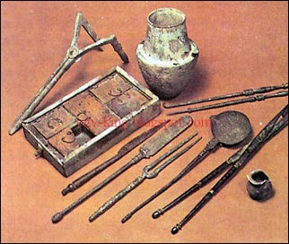
ancient Egyptian medical tools
In a study published in PLOS ONE journal in February 2023, archaeologists announced the discovery of a man who lived 3,500 years who underwent trephination and maybe had leprosy. Business Insider reported: During an archeological excavation of a tomb in Tel Megiddo, Israel, researchers discovered the remains of two Bronze Age brothers who lived between 1550 B.C. and 1450 B.C. Co-author of the study Israel Finkelstein said the city Megiddo was one of the "wealthiest and most cosmopolitan" in the region in 19th century B.C. [Source: Isobel van Hagen, Business Insider, February 26, 2023]
The older brother is estimated to have been between 21 and 46 years old, and the younger brother was estimated to be in his late teens or early 20s. Evidence of brain surgery — angular notched trephination — was discovered in the older brother. Rachel Kalisher, the lead author of the study and scholar at Brown University's Joukowsky Institute for Archaeology and the Ancient World, said that the brothers were likely elite or even royal members of their society — a reason they could have received the rare medical treatment.
The two bodies were found buried with fine pottery in a place where the community buried their upper class and elite. Researchers used DNA testing to determine that the two were brothers, and each had extensive health concerns. According to the Miami Herald: The brothers likely had developmental delays and physical deformities, but due to their high social status, they weren’t ostracized from their community. There was evidence of bone degradation and infectious lesions, potentially from tuberculosis or leprosy. [Source: Irene Wright, Miami Herald , February 23, 2023]
Researchers believe the surgery was completed while the man was alive. The beveling on the skull suggests that the cut was made into living bone and the depth of the cut suggests those completing the surgery were trying not to go too deep and into a living brain. But there was no evidence of healing on the bone, meaning the man likely died during or soon after the surgery. The surgery was “an unusual and high-level intervention that indicates access to the services of a trained practitioner who administered this treatment shortly before death,” according to the study. The researchers said the brother’s social status would have provided him access to such a unique treatment in the Late Bronze Age. Archaeological records of such a surgery are rare, and only five cases have been documented from the region. “Thus, Megiddo’s case is the earliest of its kind in the region by at least several centuries,” researchers said in the study.
"You have to be in a pretty dire place to have a hole cut in your head," Kalisher said in a statement. "I'm interested in what we can learn from looking across the scientific literature at every example of trephination in antiquity, comparing and contrasting the circumstances of each person who had the surgery done."
Dentistry Between 15,000 and 13,000 Years Ago
Archaeology magazine reported: A team led by Stefano Benazzi of the University of Bologna has discovered the earliest known evidence of dental work in a 14,000-year-old molar from a male skeleton found in northern Italy in 1988. Examination of the tooth with a scanning electron microscope revealed striations consistent with scratching and chipping with a sharp stone tool, apparently to remove decayed material. Enamel in the area of the cavity is worn away, suggesting the treatment occurred long before death. Benazzi believes that the likely very painful practice of removing tooth decay probably evolved from the use of wooden and bone toothpicks, many of which have been found at Paleolithic sites. [Source: Daniel Weiss, Archaeology magazine, November-December 2015]
In 2017, Benazzi’s team, near Lucca in northern Italy, found the oldest example of the practice of filling dental cavities. Using microscopic techniques, Benazzi and his team determined that two 13,000-year-old front teeth that belonged to a late Pleistocene hunter-gatherer were manipulated with a handheld tool. The Ice Age dentist drilled to remove decaying material within the pulp chambers of the teeth and replaced it with a natural antiseptic paste containing bitumen, vegetal fibers, and hair. This new evidence suggests a more sophisticated technology than previous markings that Benazzi and his colleagues found in teeth from another site in Italy, dated a thousand years earlier. Those teeth are believed to be the first known evidence of human dentistry. [Source: Marley Brown, Archaeology magazine, July-August 2017]
Dentistry Between 9,000 and 6,500 Years Ago
Dental work was done 9,000 years ago at Mehrgarh, a Neolithic villages in present-day Pakistan. Nine individuals from a sample of 300 buried in graves dating from 5500 to 7000 B.C. Had holes drilled in their molars. David Frayer, a professor of anthropology from the University of Kansas, wrote in Natural History, “This is certainly the first case of drilling a person’s teeth. But even more significant this practice lasted 1,500 years and was a tradition at this site. It wasn’t just a sporadic event.” The oldest recorded dentistry before the discovery was found in Denmark and dated to 3000 B.C. The discovery was reported in an article in Nature by Roberto Macciarelli of the University of Pontiers in France. “Four teeth show signs of decay associated with the hole, indicating that the intervention in some cases could have been therapeutic or palliative,” he said. No evidence of a filling was found but it is possible that there could have been something that decayed away.
The drilling was done to molars in both the upper and lower jaws to adults. In four of the cases the teeth appeared to have been drilled where the teeth had rotted but in the other cases no tooth rot was present. The holes were between a half millimeter and 3.5 millimeters deep. They appear to not have been done for aesthetic reason because the holes way out of view. The drilling is believed to have been performed with a flint point spun with a bow. An experimental reconstruction of the probable method involved a small thin piece of flint attached to a bone. Judging from the angle of the holes they were not self made and because the people who had the dental work were performed on them were not buried in special graves it appears that dentistry was available to anyone and was not just the provenance of the rich. One of the individuals had three molars drilled. Another had one molar drilled twice.
Zach Zorich wrote in Archaeology: In 2012, scientists announced that had uncovered evidence of a couple of instances of ingenious dental work in the ancient world. A team led by Federico Bernardini of the International Center for Theoretical Physics in Trieste, Italy, used a variety of techniques including CT scans and mass spectrometry to show that a 6,500-year-old skull found at the site of Lonche in Slovenia contains a cracked tooth that had been filled with beeswax—the oldest dental filling yet discovered (below, on left). A similarly inventive technique was used on an Egyptian man whose mummified body dates to around 2,100 years ago. Andrew Wade of the University of Western Ontario led a group of researchers who found that the man had numerous cavities, the largest of which had been packed with linen.” [Source: Zach Zorich, Archaeology, December 19, 2012]
Surgery in Mesopotamia

Gulam the Sumerian deity of healing
Nancy Demand of Indiana University wrote: “Among Hammurabi's laws were several that pertained to the liability of physicians who performed surgery. These laws state that a doctor was to be held responsible for surgical errors and failures. Since the laws only mention liability in connection with "the use of a knife," it can be assumed that doctors in Hammurabi's kingdom were not liable for any non-surgical mistakes or failed attempts to cure an ailment. [Source: The Asclepion, Prof.Nancy Demand, Indiana University - Bloomington +++]
“It is also interesting to note that according to these laws, both the successful surgeon's compensation and the failed surgeon's liability were determined by the status of his patient. Therefore, if a surgeon operated and saved the life of a person of high status, the patient was to pay ten shekels of silver. If the surgeon saved the life of a slave, he only received two shekels. However, if a person of high status died as a result of surgery, the surgeon risked having his hand cut off. While if a slave died from receiving surgical treatment, the surgeon only had to pay to replace the slave. This use of status to evaluate misdeeds does not seem to appear in other, similar "codes" however. +++
“Regardless of the risks associated with performing surgery, at least four clay tablets have survived that describe a specific surgical procedure. Unfortunately, one of the four tablets is too fragmentary to be deciphered. Of the remaining three, one seems to describe a procedure in which the asu cuts into the chest of the patient in order to drain pus from the pleura. The other two surgical texts belong to the collection of tablets entitled "Prescriptions for Diseases of the Head." One of these texts mentions the knife of the asu scraping the skull of the patient. The final surgical tablet mentions the postoperative care of a surgical wound. This tablet recommends the application of a dressing consisting mainly of sesame oil, which acted as an anti-bacterial agent.” +++
See Separate Article: HEALTH, ILLNESSES AND MEDICINE IN MESOPOTAMIA africame.factsanddetails.com
Surgery in Ancient Egypt
Some surgery was done. Skulls with holes in them have been found. Surgical tools used by a Egyptian doctors included: knives, adrill, saw, forceps or pincers, censer, hooks, bags tied with string, beaked vessel, vase with burning incense, Horus eyes, scales, pot with flowers of Upper and Lower Egypt, pot on pedestal, graduated cubit or papyrus, scroll without side knot (or a case holding reed scalpels), shears, spoons. ~
The Temple of Sobek and Horus in Kom Ombo in Aswan contains sculpted wall reliefs with images of surgical instruments, bone saws and dental tools. The Cairo Museum has a collection of surgical instruments which include scalpels, scissors, copper needles, forceps, spoons, lancets, hooks, probes and pincers. [Source: Mark Millmore,discoveringegypt.com discoveringegypt.com ^^^]
According to Minnesota State University, Mankato: “Though Egyptian medical practices by no means could rival that of the present day physicians, Egyptian healers engaged in surgery, prescriptive, and many other healing practices still found today.The practices of Egyptian medical practitioners ranged from embalming to faith healing to surgery and autopsy. The use of autopsy came through the extensive embalming practices of the Egyptians, as it was not unlikely for an embalmer to examine the body for a cause of the illness which caused death. The use of surgery also evolved from a knowledge of the basic anatomy and embalming practices of the Egyptians. From such careful observations made by the early medical practitioners of Egypt, healing practices began to center upon both the religious rituals and the lives of the ancient Egyptians. [Source: Minnesota State University, Mankato, ethanholman.com]
Egyptians used splints to set broken bones. Anatomical knowledge appears to have been based more on animals than humans. Bodies were believed to have been cut open with obsidian blades. Bronze and copper knives were not sharp enough. In lieu of anesthesia patients were perhaps knocked on the head. The oldest set of bronze surgical blades dates to 2300 B.C.
See Separate Article: HEALTH CARE IN ANCIENT EGYPT factsanddetails.com
Surgery in Ancient Greece

votive releif
A set of Greek medical instruments consisted of catheters, a rectal speculum, scoops, probes, hooks, forceps, traction hooks and bone chisels. Cauterizing (burning of part of a body to remove or close off a part of it) was a standard medical procedure in ancient times. Brain-swelling was sometimes relived with trephination, an ancient medical technique in which holes were cut into the brain to relieve pressure. It was procedure thought to be only performed on the elite.
Broken bones, surprisingly, were often not set, meaning that victims were disfigured for the rest of the lives. Other times bones were set with skill. "This was a remarkable surgical procedure," an archaeologist told National Geographic, displaying a thigh bone from an ancient Greek skeleton. "Someone used a lot of force to pull the lower part of the broken bone down, reset it, and keep it in place for weeks against the enormous pressures of contracting muscles."
There were several factors that influenced the development of medicine in ancient Greece. First, there was the potent force of religion with its gods and goddesses who dealt with healing, death and pestilence. Then there was the influence of trading contacts such as Egypt (which had learned much from its mummification practices) and Mesopotamia (which had published comprehensive medical documents on clay tablets well before 1000 B.C.). [Source: Canadian Museum of History |]
To cap it off, there was the sad result of war - a variety of wounds and amputations caused by arrows, swords, spears and accidents- and described so vividly and accurately in Homer's Iliad. Just dealing with these casualties provided lots of experience and practical information applicable elsewhere. Although Greek religion frowned on human dissection in the Archaic and Classical periods, after the founding of the Alexandrian School that changed. Physicians and researchers made advances in some areas that were not surpassed until the 18th Century.” |
See Separate Article: SURGERY IN ANCIENT GREECE AND ROME europe.factsanddetails.com
Image Sources: Wikimedia Commons except Borneo amputation from Tim Maloney,
Text Sources: Internet Ancient History Sourcebook: Rome sourcebooks.fordham.edu ; Internet Ancient History Sourcebook: Late Antiquity sourcebooks.fordham.edu ; “Outlines of Roman History” by William C. Morey, Ph.D., D.C.L. New York, American Book Company (1901) ; “The Private Life of the Romans” by Harold Whetstone Johnston, Revised by Mary Johnston, Scott, Foresman and Company (1903, 1932); BBC Ancient Rome bbc.co.uk/history/ ; Project Gutenberg gutenberg.org ; Metropolitan Museum of Art, National Geographic, Smithsonian magazine, New York Times, Washington Post, Los Angeles Times, Live Science, Discover magazine, Archaeology magazine, Reuters, Associated Press, The Guardian, AFP, The New Yorker, Wikipedia, Encyclopædia Britannica, Encyclopedia.com and various other books, websites and publications.
Last updated November 2024