BIOTECHNOLOGY ADVANCES IN JAPAN
Japanese researchers have worked out ways to extract rubber from mushrooms, treat cancer with chemicals from bioluminescent jellyfish and created mice that glow in the dark and frogs with transparent skin whose internal organs can studies while they are functioning.
Engineers at the Shimadzu Corporation developed mass spectroscope capable of identifying and analyzing “biological macromolecules” through vaporizing samples and study the speed and dispersion of the main component. The machine was widely regarded as the best of its kind in the world but only one was sold: to Koichi Tanaka who found it indispensable in his research that won him a Nobel Prize in chemistry in 2002.
In May 2001, two Japanese scientists were charged with stealing genetic material relating to Alzheimer's research. It was the first time anyone had been arrested in the United States under the Economic Espionage Act of 1996. The Japanese scientists allegedly stole vials of DNA samples and left behind vials with tap water.
In 2008, a team at Tokyo University was able to produce healthy mice offspring without male participation, using “bi-maternal embryos” that only involve female genomes. The success rate was 30 percent about the same rate as in-vitro fertilization or normal embryos with male sperm. Some genetically modified female grew into adults that produce their own offspring.
Scientists at Mie University and Nagoya University have developed see-through goldfish with transparent scales in their skin. The scientists said the advance was more than a curiosity and will help scientist research blood constituents and organ behavior.
Tokyo University of Science researchers have implanted bio engineered seedlike tissue into the jaws of mice that grew into new teeth. It is hoped the technology, published in the Proceedings of the National Academy if Sciences, can be used not only to grow teeth but also to grow organs in humans.
Medical Advances in Japan

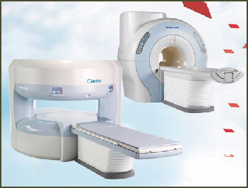
Hitachi MRI Doctors at Osaka University have succeeded in restoring eyesight to blind patients by transplanting corneas created by cultivating cells from patients oral mucous membranes, which contain many stem cells which can be used to create certain tissues in the cornea.
In January 2007, a team of researchers with the National Cancer Center Research Institute and the International Medical Center of Japan announced that they had created a liver cell from a fat cell. The development hold promise for treating liver diseases such as hepatitis and cirrhosis and could be used to create cell for other organs from fat.
A team at Waseda University lead by biochemist Eishun Tsuchida has developed completely synthetic blood. The only problem at this juncture is that it costs about $100,000 a liter to make. The secret is synthesizing the protein albumin using yeast as the basic material and as an alternative to hemoglobin. It is hoped that synthetic blood can one day replace real blood and has the advantage in that it can be used by people of all blood types. The Waseda team is currently working on developing methods to reduce the price of production.
Japanese doctors use weak ultrasonic waves to hasten the healing of broken bones. The process shortens the recover period of broken femurs and wrist by about 40 percent and is sometimes uses instead of surgery. It is still not clear why recovery is so rapid. The frequency used is the same as that used in ultrasound test of fetuses.
The Japanese have developed custom-made bones that can be instantly made from calcium phosphate — the substance of which real bones are made — and put on the place of damaged bones after an accident. The technology is current in the clinical trials stage. Doctors usually mend damaged bones with transplanted bones or ceramic substitutes.
Nakanishi Inc. in Tanuma, Tochigi, is a world leader in the making of dental drills. Controlling 20 percent of the market, vying for the No. 1 position with a German rival, it produces drills that can saw away decay at 1 million rotations every 2½ minutes and weighs less than 100 grams.
Stem Cell Research in Japan
Japanese scientists have made a number of breakthroughs with stem cells. In May 2007, scientists at Kobe University succeeded in mass cultivating of embryonic stem cells and developed them into brain cells. In 2008 researchers at the Institute of Medical Science at Tokyo University announced the creation of mouse pancreases and kidneys from embryonic stem cells.
In January 2011, Japanese scientists at Tottori University said they had successfully used stem cells to successfully make heart pacemaker cells in mice, an advancement that could lead to a breakthrough in the treatment of irregular heartbeats and heart diseases in humans.
Researcher at the Kyushu University and Osaka University are trying to use stem cells from mastectomy patients to help them regrow their breasts.
Stem Cell Heart Treatment
A team lead by Hiroaki Matsubara of Kyoto Prefectural University of Medicine announced in July 2010, it had harvested stem cells from a patient’s heart and used them grow new cells which were injected into necrotic tissue in the heart to regenerate the muscle. The treatment has been used to help people who have suffered heart attacks and is being touted as an alternative to heart transplants.
A patient named Shideki Yamaguchi from Kobe with heart disease — who suffered a heart attack and was considered beyond hope for bypass testaments — underwent the treatment and recovered enough to resume his daily life activities without assistance. Before the procedure he had been bed-bound.
In Yamaguchi’s cases 15 milligrams of coronary tissues was harvested by inserting a thin tube into a blood vessel running from his groin to his heart. Stem cells were cultivated for a month and their numbers multiplied about 40,000 times. The cells were injected during bypass surgery to an area of the heart where cells had been dying because of lack of blood flow.
Automatic IPS culture has been achieved it was announced in June 2010, by Kawasaki Industries and two state-backed laboratories — KHL and AIST. The breakthrough hold the potential of mass-producing IPD cells which would have a host of applications.
Immune-deficient Pigs Created for Organ Transplant Research
In June 2012, Jiji Press reported: “A team of Japanese scientists has succeeded in creating an unprecedented type of immune-deficient pig that can be used for regenerative medicine research and the development of new drugs. The male pigs, created by combining genetic engineering and cloning techniques, lack a thymus (an immune organ) and have no T cells or natural killer (NK) cells, both lymphocytes, according to the team that includes researchers from the National Institute of Agrobiological Sciences and Riken. In addition, the pigs' B cells, also lymphocytes, are unable to produce antibodies. The team announced the creation of the pigs in U.S. magazine Cell Stem Cell's edition. [Source: Jiji Press, June 18, 2012]
Next, the team plans to produce human organs in the bodies of the immune-deficient pigs using human induced pluripotent stem (iPS) cells and embryonic stem (ES) cells, and evaluate the organs' safety and efficiency. In addition, the effects and side effects of potential new drugs will be analyzed using the pigs. In the future, the team plans to create pigs that cannot produce B cells, and cross them with immune-deficient pigs to establish a more severely immunodeficient strain.
“The pigs created by the team can live for a long time if they are raised in a biological clean room or undergo bone marrow transplants. They will die at the age of 2 months if bred in closed facilities for genetically modified animals. According to the team, immune-deficient male pigs can reliably be produced if normal male pigs are bred with potentially immune-deficient female pigs created with the same techniques as those used to develop the pigs in the study.
Cell Sheets

Yoshio Yamada
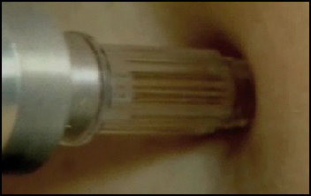
needle free injection A medical team led by Prof. Yoshiki Sawa at Osaka University succeeded in restoring functions to the diseased heart of a patient with severe cardiac arrest using muscle cells taken from the patient’s thighs. The male patient, in his 50s, had been waiting for a heart transplant. It was the first time that a person waiting for an organ transplant was treated using his own cells.
The treatment involved removing about 10 grams of muscles tissue from the patient’s thigh. From the muscle tissue the doctors extracted myobalst cells, which are the main building blocks for muscle fibers. The team cultivated the cells and formed them into sheets about four centimeters wide and 0.05 millimeters thick and then wrapped he diseased heart with three layers of myobalst cells.
The team spent two months creating the myoblast sheets. They were removed from the patient on March 2007 and attached to heart, mainly around the heart’s left ventricle, in May. After the treatment the pulse rate and quality of blood pumped from the heart all improved. Three months after the treatment the patient was able to leave the hospital on foot without a pacemaker he had before the treatment.
Prof. Sawa said, “The myobalst sheets were not transformed into heart muscle, but they apparently released substances that assist the functioning of weakened heart muscles.”
Cloning in Japan
In July 1998, Japanese scientists announced that they had created two calves cloned from an adult cow. The calves were the first animals cloned from an adult since Dolly, the sheep, was cloned about 17 months earlier. Later a 2nd generation cloned calf was produced.
Japanese postdoctoral student Teruhuko Wakayama figured out a better way to clone mammals than the Dolly method. In 1998 at the University of Hawaii he cloned mice with a 2 out of 100 success rate (compared to 1 to 277 with the Dolly method) in which a nucleus is sucked out of a cell and injected into an empty egg and stimulated with a chemical bath.
In November 2008, scientists at the Institute of Physical and Chemical Science (RIKEN) announced they had succeeded into cloning using tissue from a mouse that as frozen for 16 years, It was the first animal in the world to have been be cloned from frozen animal tissue. The achievement was done nay taking the nucleus of cells from the frozen mouse lacing them in cells of a healthy mouse that were transferred to womb of a surrogate mouse,
Reproducing a Wooly Mammoth
Kagoshima University professor Kazufumi Goto believes that is possible to produce a living wooly mammoth by: 1) breeding a mammoth (using its sperm from a mammoth to fertilize the egg of Asian elephant and repeatedly breeding the offspring to get an animal closer and closer to a mammoth; and 2) cloning a mammoth using DNA taken from a part of a mammoth and fusing it in the egg of an Asia elephant that has been stripped of its elephant genes so the baby would be a mammoth not a hybrid.
In 1990, Goto led a team that produced a healthy calf using the sperm from a dead bull. In 1996, Goto began his search for male mammoths with sperm-fill reproductive organs near Yakutsk, Siberia, where numerous frozen mammoths have been found.
The chance of finding DNA intact in frozen mammoth sperm is still very remote. Goto has offer $10,000 for mammoth tissue with intact DNA. If a viable embryo is produced it will shipped after five cell divisions to a lab in Thailand at Mahidol university, which has successfully fertilized Asian elephant eggs in vitro. The embryo would then be implanted in a surrogate and ideally emerge as a wooly mammoth 600 days later.
In the early 2000s, scientists at Kinki University in Wakayama tried to clone a mammoth using skin, leg muscle tissue and bone from a mammoth found 1,200 kilometers north of Yakutsk in Siberia.
New research announced in the late 1990s suggest that the idea of bringing a wooly mammoth back to life may be not as far-fetched as once thought. About 80 percent of the mammoth genome has been pieced together from samples taken from two carcasses found in Siberia. Among the discoveries related to this is that mammoth are much more closely related to modern elephants than previously thought.
The biggest obstacle to overcome will be getting a mammoth cell into good enough shape to inject into a an egg. In most cloning cases a cell is taken from a live animal and injected into the egg. Obviously things are different with a cell taken from a mammoth carcass that as been sitting around for 10,000 years. Instead do being neatly arranged the chromosome are in little pieces and they will have to be reconstructed, something that is far beyond the reach of today’s science.
Scientist at Tokyo’s Institute of Technology have dissected a coelacanth in.
Latest on Resurrecting a Mammoth in Japan
In January 2011 it was announced that a team of researchers would make a serious attempt to bring back to life a mammoth species using new cloning technologies after obtaining tissue from the carcass of a mammoth preserved in a Russian mammoth research laboratory. "Preparations to realize this goal have been made," Prof. Akira Iritani, leader of the team and a professor emeritus of Kyoto University, told the Yomiuri Shimbun. [Source: Yomiuri Shimbun, January 13, 2011]
The Yomiuri Shimbun, “Under the plan, the nuclei of mammoth cells will be inserted into an elephant's egg cells from which the nuclei have been removed to create an embryo containing mammoth genes. The embryo will then be inserted into an elephant's womb in the hope that the animal will give birth to a baby mammoth.”
Researchers from Kinki University's Graduate School of Biology-Oriented Science and Technology began the study in 1997. On three occasions, the team obtained mammoth skin and muscle tissue excavated in good condition from the permafrost in Siberia.However, most nuclei in the cells were damaged by ice crystals and were unusable. The plan to clone a mammoth was abandoned.
In 2008, Dr. Teruhiko Wakayama of Kobe's Riken Center for Developmental Biology succeeded in cloning a mouse from the cells of mouse that had been kept in deep-freeze for 16 years. The achievement was the first in the world. Based on Wakayama's techniques, Iritani's team devised a technique to extract the nuclei of eggs — only 2 percent to 3 percent are in good condition — without damaging them.
In the spring of 2010, the team invited Minoru Miyashita, a professor of Kinki University who was once head of Osaka's Tennoji Zoo, to participate in the project. He asked zoos across the nation to donate elephant egg cells when their female elephants died. The team also invited the head of the Russian mammoth research laboratory and two U.S. African elephant researchers as guest professors to the university. The research became a joint effort by Japan, Russia and the United States.
If a cloned mammoth embryo can be created, Miyashita and the U.S. researchers, who are experts in animal in vitro fertilization, will be responsible for transplanting the embryo into an African elephant. The team told the Yomiuri Shimbun if everything goes as planned, a mammoth will be born in five to six years. "If a cloned embryo can be created, we need to discuss, before transplanting it into the womb, how to breed [the mammoth] and whether to display it to the public," Iritani said. "After the mammoth is born, we'll examine its ecology and genes to study why the species became extinct and other factors."
Scientists a Step Closer to Cloning Mammoth
In December 2011, Kyodo reported: “The thighbone of a mammoth found in August in Siberia contains well-preserved marrow, increasing the chances of cloning one of the extinct beasts, Japanese and Russian scientists confirmed recently. The teams from the Sakha Republic's mammoth museum in eastern Russia and Kinki University's graduate school in biology-oriented science and technology will launch full-fledged joint research next year to clone the giant mammal, which is believed to have become extinct about 10,000 years ago, they said. [Source: Kyodo, December 4, 2011]
“By transplanting nuclei taken from the marrow cells into elephant egg cells whose nuclei have been removed through a cloning technique, embryos with a mammoth gene could be produced and planted into elephant wombs, as the two species are close relatives, they said. Securing nuclei with an undamaged gene is essential for the nucleus transplantation technique, but doing so from mammoths is extremely difficult and scientists have been trying to reproduce a mammoth since the late 1990s, they said.
“In the Sakha Republic, global warming has thawed its almost permanently frozen ground, leading to numerous discoveries of frozen mammoths. But cell nuclei are usually damaged or have not been kept in a frozen state even when they have been found in a good overall condition, a Russian museum official said. This time, however, there is a high likelihood that biologically active nuclei can be extracted as the frozen marrow found when museum scientists cut open the thighbone Nov. 13 was fresh and in excellent condition, according to the official. The bone was found near Batagay in northern Sakha.
“The technique for extracting nuclei, meanwhile, has improved dramatically in the past few years and some undamaged nuclei have been successfully taken from badly preserved mammoth tissue fragments, albeit at low rates, said the Kinki University team based in Osaka Prefecture.
“The museum, located in the republic's capital, Yakutsk, soon notified the Japanese side, with which it has had close ties through joint research since 1997, including professor Akira Iritani and associate professor Hiromi Kato. Iritani confirmed that the outstanding condition of the marrow has increased the chances of cloning a mammoth, and said the Japanese team will try to obtain elephant eggs for the research project, although he added this would not be easy.
Unlocking the Secret of Life at Tokyo University
A group of scientists at the University of Tokyo has created a protocell capable of self-replicating, a feat that may provide clues to understanding how life was created and multiplied, according to the group. The scientists announced their achievement in the online edition of British scientific journal Nature Chemistry. [Source: Yomiuri Shimbun, September 8, 2011]
The scientists created a cell membrane measuring one-hundredth of a millimeter in diameter that formed an artificial cell containing a fluorescent protein-producing gene. They then utilized changes in temperature and other factors to cause DNA strands in the cell to multiply. Adding cell membrane material to the cell caused it to divide, the group said.
The group determined how much DNA had been replicated by studying the amounts of luminescence generated by the artificial protocell. Their results showed that the faster the DNA strands in a protocell multiplied, the faster that cell would divide and multiply.Some protocells divided three to four times within 10 minutes; the multiplied DNA strands in those cells are believed to have stimulated division-triggering membranes, according to the group.
"In the creation of life, cell membranes might have been created first, with gene-like substances penetrating the membranes to cause cellular division," Tadashi Sugawara, a professor emeritus at the university, said. "In my future research, I want to create an artificial cell that continues to divide and multiply with little artificial manipulation."
Image Sources:
Text Sources: New York Times, Washington Post, Los Angeles Times, Times of London, Yomiuri Shimbun, Daily Yomiuri, Japan Times, Mainichi Shimbun, The Guardian, National Geographic, The New Yorker, Time, Newsweek, Reuters, AP, Lonely Planet Guides, Compton’s Encyclopedia and various books and other publications.
Last updated October 2012
